Cell Biology
III. DNA and Protein Synthesis
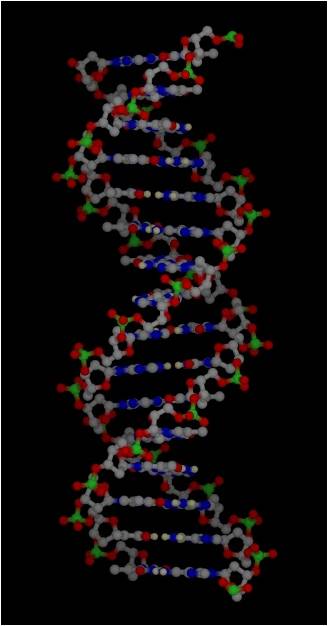 Much of the energy harvested by a cell is used to make proteins. However, in order to understand protein synthesis, we must first describe the structure of DNA. DNA is the 'recipe' for proteins, so we need to take a little detour on our jounrey of how a cell works to describe the structure of DNA and chromosomes. DNA
is the genetic material in all forms of life (eubacteria, archaea, protists,
plants, fungi, and animals). Those quasi-living viruses vary in their genetic
material. Some have double-stranded DNA (ds-DNA) like living systems, while
others have ss-DNA, ss-RNA, and ds-RNA. RNA performs a wide array of functions
in living systems. Many of these functions have only been discovered in the
last few years.
 A.
DNA and RNA Structure
DNA (deoxyribonucleic acid) and RNA
(ribonucleic acid) are nucleic acids - polymers consisting of a linear
sequence of linked nucleotide monomers. We will describe the structure of the
monomers first, and then describe how they are linked into linear polymers.
Finally, we will describe the double-stranded structure of ds-DNA.
1. The monomers
are "nucleotides"
three components:
 -
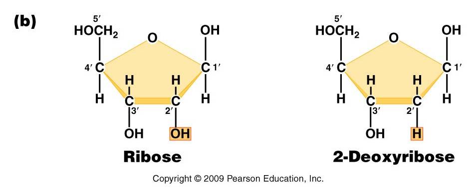
Pentose (5 carbon) sugar: either ribose (RNA) or deoxyribose
(DNA). The carbons are numbered clockwise. The difference between the sugars
is that ribose has an -OH group on the 2' carbon, whereas deoxyriboes has
only 2 H groups and thus is "deoxygenated" relative to ribose. BOTH
sugars have an -OH group on the 3' carbon, which will be involved in binding.
The 5' carbon is a sidegroup off the ring.
-
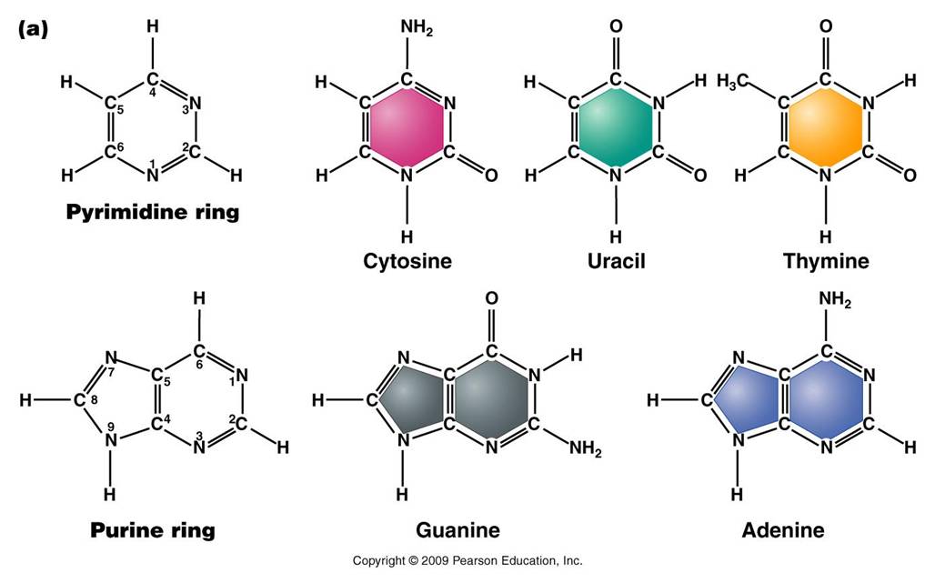
Nitrogenous Base: each nucleotide has a single nitrogenous
base attached to the 1' carbon of the sugar. This nitrogenous base may be
a double-ringed structure (purine) or a single ringed (pyrimidine) structure.
The purines are adenine (A) and guanine (G). The pyrimidines are thymine (T),
cytosine (C), and uracil (U). DNA nucleotides may carry A, G, C, or T. RNA
nucleotides carry either A, G, C, or U.
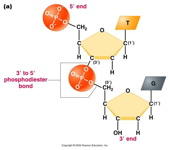 -
The third component of a nucleotide is a phosphate group,
which is attached to the 5' carbon of the sugar. When a nucleotide is incorporated
into a chain, it has a single phosphate group. However, nucleotides can occur
that have two or three phosphate groups (dinucleotides and trinucleotides).
ADP and ATP are important examples of these types of molecules. In fact, the
precursors of incorporated nucleotides are trinucleotides. When two phosphates
are cleaved, energy is released that can be used to add the remaining monophosphate
nucleotide to the nucleic acid chain.
2. Polymerization
is by 'dehydration synthesis'
As with all other classes of biologically
important polymers, monomers are linked into polymers by dehydration synthesis.
In nucleic acid formation, this involves binding the phosphate group of one
nucleotide to the -OH group on the 3' carbon of the existing chain. For the
purposes of seeing how this reaction works, we can envision an H+ on one of
the negatively charged oxygens of the phosphate group. Then, a molceule of water
can be removed from these two -OH groups, leaving an oxygen binding the sugar
of one nucleotide to the phosphate of the next.
This creates a 'dinucleotide'. It
has a polarity/directionality; it is different at its ends. At one end, the
reactive group is the phosophate on the 5' carbon. This is called the 5' end
of the chain. At the other end, the reactive group is the free -OH on the 3'
carbon; this is the 3' end of the chain. So, a nucleic acid strand has a 5'
- 3' polarity.
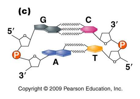 3.
Most DNA exists as a 'double helix' (ds-DNA) containing two linear nucleic acid
chains.
a. the
nitrogenous bases on the two strands are 'complementary' to each other,
and form weak hydrogen bonds between them. A always pairs with T, and C always
pairs with G. As such, there is always a double-ringed purine pairing with
a single-ringed pyrimidine, and the width of the double-helix is constant
over its entire length.
b. the
two strands (helices) are anti-parallel:
they are arranged with opposite polarity. One strands points 5' - 3', while
the other points 3' - 5'. The direction of the pentose sugars and the type
of reactive group at the ends of the chains show this relationship.
4. RNA performs
a wide variety of functions in living cells:
a. m-RNA
(for "messenger") is the copy of
a gene. It is the
sequence of nitrogenous bases in m-RNA that is actually read by the ribosome
to determine the structure of a protein.
b. r-RNA
(for "ribosomal")
is made the same way, as a copy of DNA. However, it is not carrying the recipe
for a protein; rather, it is functional as RNA. It is placed IN the Ribosome,
and it helps to ‘read’ the m-RNA.
c. t-RNA
(for "transfer")
is also made as a copy of DNA, but it is also functional as an RNA molecule.
Its function is to bind to a specific amino acid and incorporate it into the
amino acid sequence as instructed by the m-RNA and ribosome.
d. mi-RNA
(micro-RNA) and si-RNA (small interfering RNA)
bind to m-RNA and splice it; inhibiting the synthesis of its protein. This
is a regulatory function.
e.
sn-RNA (small nuclear RNA) are short sequences that process
initial m-RNA products, and also regulate the production of r-RNA, maintain
telomeres, and regulate the action of transcription factors. Regulatory functions.
B. Protein Synthesis
As we've already
mentioned, protein synthesis is fundamental to nearly everything a cell does.
Protein channels are used to transport large molecules across the membrane.
Almost all chemical reactions occuring in cells are catalyzed by protenaceous
enzymes, including those involved in energy harvest, DNA replication, and cell
division. Proteins perform important structural functions within cells and multicellular
organisms, too; such as the histone proteins in chromosomes, the proteins in
ribosomes, the collagen and elastin fibers that hold skin cells together, the
collagen on which calcium and phosphate is deposited in bone, the protein myofibrils
of actin and myosin in muscle cells, the neurotransmitters used for cell-cell
communication between neurons, and the enzymes that digest food in the stomach
and intestine of animals. So, proteins are fundamental to what cells and organisms
ARE, structurally, and what they DO functionally. As you know, the genetic information
determines the types of proteins a cell can make. The subset of proteins a cell
actually DOES make, and the timing of WHEN they are made, is determined by what
genes are "on" and what genes are "off" at a given time.
This regulation of gene activity is ALSO co-ordinated by proteins - called transcription
factors - that bind to DNA and promote or inhibit gene activity. So, proteins
also regulate protein synthesis. Hopefully you see just how important proteins
are to cells and organisms. So, the process of making these proteins is important,
too. It is no surprize, then, that a lot of the energy harvested by a cell is used to make proteins. The phosphate bonds in ATP are broken, and the energy that is released is used to form new bonds between amino acids. The chain of amino acids that is produced becomes a functional protein.
 1.
Overview
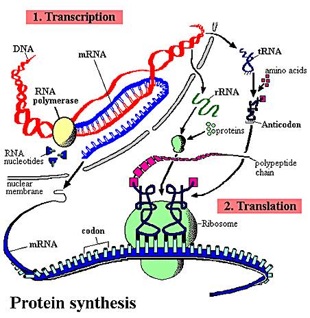
The sequence of nitrogenous
bases in a region of DNA is 'read' by a complex of enzymes that build a complementary
strand of RNA. This process of reading DNA and making RNA is called 'transcription'.
This is a great word for the process, as the message written in the language
of nucleic acids is copied in essentially the same language - the language of
nucleic acids. This RNA may be a recipe for a protein (m-RNA), or it may be
an RNA that will act on its own as t-RNA, mi-RNA, si-RNA, or be complexed with
proteins in the ribosome (r-RNA). Obviously, in "protein synthesis",
only the m-RNA is read to make a protein. However, the other molecules all play
a role. The sequence of nitrogenous bases in the m-RNA is then 'read' by a ribosome,
which links a specific sequence of amino acids together into a protein based
on that sequence of nitrogenous bases in the m-RNA. This process is called 'translation'.
This is a great choice of a word, too. Here the sequence of information written
in the language of nucleic acids is rewritten in a new language (hence, translation)
- the language of amino acids.
Many of the initial RNA products
have specific regions (introns) cut out of their sequence before they become
functional. This step is known as "RNA processing" or "RNA splicing".
Introns are present in nearly all eukaryotic RNA's, and are also in the DNA
genes that encode them. Up until a few years ago, the only introns in prokaryotes
had been found in t-RNA molecules of archaeans. More recently, however, introns
have been found in m-RNA and r-RNA molecules of a few eubacteria and a few more
archaeans. So, although they are rare in prokaryotes, we will describe a generic,
simplified process of protein synthesis that includes introns and RNA processing.
In addition to splicing the RNA product
of transcription, the initial protein product of translation may also be spliced
and modified before it becomes functional. In eukaryotes, this protein processing
often occurs in the Golgi apparatus.
The description presented here is
a simple model of protein synthesis. You will learn more complex aspects of
this process in Genetics.
2. The Process of Protein Synthesis
1. Transcription:
 a. The message is on one strand of the
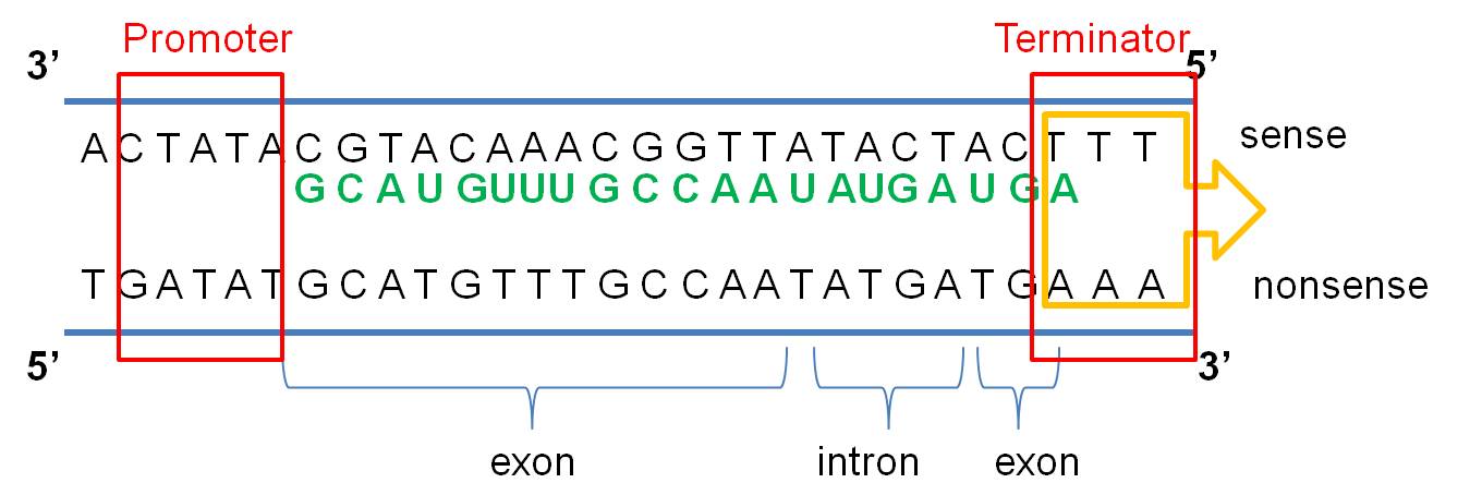
double helix - the sense strand: The DNA double helix is composed
of two anti-parallel complementary strands of DNA. Only one strand in a coding
region ("gene") is read; this strand carries a meaningful recipe that
"makes sense". This is called the "sense" strand. The other
strand, limited by complementarity, is not a meaningful message - it is the
"non-sense" (or "anti-sense") strand. Think about it this
way. Given a meaningful message of "C-A-T" (a small furry mammal),
the complementary strand is limited to the meaningless sequence of "G-T-A"
(????...). As the meaningful sequence gets longer, it is even LESS likely that,
just by chance, the complementary strand would be meaningful, too. Again, in
all eukaryotic genes and in some rare prokaryotic ones, there will be non-functional
"introns" interspersed throughout the meaningful message. The meaningful
parts are called "exons". The process of transcription is continuous,
so introns and exons get transcribed and these regions - if present in the DNA
- will also be present in the RNA product.
b.
The cell 'reads' the correct strand based on the location of the promoter, the
anti-parallel nature of the double helix, and the chemical limitations of the
'reading' enzyme, RNA Polymerase. RNA Polymerase binds to the
DNA at a specific sequence next to the gene, called the 'promoter'. It binds
in a specific way, so it is pointed towards the gene. RNA polymerase can only
create a strand of RNA in the 5' to 3' direction, adding a new base to the free
-OH group of the preceeding nucleotide on the chain. So, from its position at
the promoter, looking down the two strands in the gene, the RNA Polymerase can
only 'read' one strand - the DNA strand that is 3'-5'. It must create a strand
that is anti-parallel to the DNA' template', and it can only bind nucleotides
in 5'-3' direction. So, only the 3'-5' DNA strand is read in this region, and
only one RNA strand, 5'-3' is made. It is important to appreciate that this
relationship is 'local'. In another region of the DNA, the promoter may be on
the other side of the gene, and the other strand may be read.
c.
Transcription ends at a sequence called the 'terminator'.These
regions have specific sequences that destabilize the attachment of the RNA Polymerase
to the DNA... it detaches and transcription stops. VIDEO
So, the process of transcription can be summarized like this: RNA Polymerase
binds at the promoter and reads the sense strand of DNA. The ploymerase links
together RNA nucleotides 5--> 3, in a sequence complementary to the DNA sense
strand. This process is continuous, so all DNA bases are 'read', including exon
and intron sequnces. This process continues until a terminator region is reached.
Reading this region destabilizes the RNA polymerase. It detaches from the DNA,
and transcription stops. All types of RNA (m-RNA, r-RNA, t-RNA) are made through
this process.
 2.
Transcript Processing:
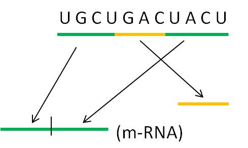
At this point in the process, the
cell has read the gene and synthesized a complementary copy of strand of RNA.
In all eukaryote sequences and many prokaryotic ones, this RNA molecule will
have non-functional introns that need to be 'cut-out'. Enzymes cut the introns
out and splice the ends together. In some cases, the introns catalyze their
own excision - they are RNA molecules with enzymatic activity. These are one
class of "ribozymes" - a very interesting class of molecules. There
are other ribozymes that cleave other RNA molecules (not themselves) and others
that catalyze other chemical reactions unrelated to RNA splicing.
In eukaryotes, the m-RNA, t-RNA and
r-RNA is shunted through the nuclear membrane to the cytoplasm. In prokaryotes,
there is no nucleus so the RNA is already in the cytoplasm. In all organisms,
the r-RNA is complexed with proteins to form functional ribosomes. The t-RNA's
bind specific amino acids.
VIDEO
3. Translation:
In this process,
amino acids are linked together into a protein. The particular sequence of amino
acids that are linked together is determined by the sequence of nitrogenous
bases in m-RNA. This process occurs at the ribosome.
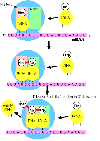 a.
m-RNA attaches to the ribosome at the 5' end. The ribosome has two reactive sites. The RNA
moves through the ribosome until a specific sequence of nucleotides, AUG, is
positioned in the first site. Three base sequence in the m-RNA are called 'codons'.
This specific codon AUG, which starts the process of translation, is called
the 'start codon'. All proteins made by all life forms initially begin with
methionine, and use the codon AUG..
b. a specific
t-RNA molecule, with a complementary UAC anti-codon sequence, binds to the m-RNA/ribosome
complex. This t-RNA
always carries the amino acid methionine. The genetic code describes the relationship
between 3-base codons in m-RNA and the amino acids they code for.
c. Binding
of the t-RNA to the first site opens a second site that reads the second 3-base
codon (GCC in picture at right). Another
t-RNA binds here - one with the specific anti-codon sequence (CGG). This t-RNA,
with this anti-codon, always binds with the amino acid alanine.
d. Now a
complex series of reactions occurs. Methionine is cleaved from its t-RNA and bound to alanine (this peptide bond
between amino acids forms via dehydration synthesis). The t-RNA in position
1 vacates the site, and the t-RNA in site 2 moves to site 1. This is called
a 'translocation reaction'. The next 3-base codon is positioned in the second
site - ready to accept the next t-RNA/ammino acid complex (for tryptophan in
the picture to the right).
e. Polymerization
proceeds. This process
continues down the m-RNA strand, reading the message one codon (3-base "word")
at a time. For each codon, a specific amino acid is added to the chain. Thus,
the nucleotide sequence in the m-RNA - copied from the nucleotide sequence in
the DNA gene - determines the sequence of amino acids in the protein.
f. Termination. There are some codons that have no corresponding t-RNA molecule. When these
codons enter the second site, no t-RNA/amino acid is added. When the ribosome
translocates, no new amino acid is added and the chain is terminated. These
particular codons that stop translation are called "stop codons".
VIDEO
4. Protein Processing:
The initial protein product usually
needs to be modified to become functional. These modifications are termed "post-translational
modifications". First of all, the methionine is usually cut off - this
relieves an important constraint on the structure of functional proteins...
functional proteins DON'T all start with methionine! Then, the protein may be
spliced, or it may be bound with a sugar group (glycoprotein), lipid (lipoprotein),
nucleic acid (nucleoprotein), or another protein (quaternary protein). In eukaryotes,
much of this processing occurs in the Golgi apparatus.
3. Regulation
of Protein Synthesis
Aside from somatic mutations, all
the cells in a multicellular organism are genetically identical. So, the cells
in your retina, bone, muscle, and stomach lining all contain the same genes.
These cells perform different functions because they are reading different genes
and making different proteins. Your muscle has the gene for rhodopsin (a photoreceptive
pigment produced in the retina), but that gene is not transcribed in muscle
cells. In contrast, retinal cells have the genes for the muscle proteins actin
and myosin, but these genes are not transcribed. So, cell specialization and
the developmental process by which cells specialize from the fertilized egg
occurs by regulating this process of protein synthesis. Regulation can occur
at each of the steps described above.
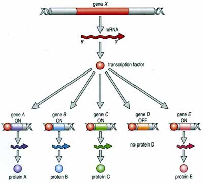 1.
Regulation of Transcription:
The
process of transcription is regulated in several ways. First, the RNA polymerase
can be blocked from the promoter. This can happen because the gene is bound
to histones in a nucleosome, or is in a region of condensed 'heterochromatin',
or because other proteins called 'transcription factors' have bound to the DNA
- either at the promoter or between it and the gene, blocking the polymerase's
route. However, the binding of other transcription factors can increase the
affinity of the RNA polymerase for the promoter - increasing the probability
of transcription. Again, these transcription factors are proteins encoded by
other genes, and affected by other cellular processes. In this way, the action
of a gene can be co-ordinated with the activity of other genes in a complex
and interdependent manner. In addition, environmental cues from outside the
cell can, through signal transduction, affect the activity of transcription
factors and turn genes on or off. So, an organism can respond genetically to
environmental cues.
2. Regulation
of Transcript Processing:
The production of a protein can be
affected at the processing stage. mi-RNA's and si-RNA's are small RNA molecules
encoded by their own genes. These molecules can bind to m-RNA and effectively
block correct splicing. This turns off production of the correct protein. In
some cases, an initial m-RNA can be spliced two ways, creating two different
functional products (and eventually two different proteins) depending on the
pattern of cleavage. So, one gne may code for different proteins in different
cells or tissue types.
3. Regulation
of Translation:
One way that differential splicing
can affect protein production is by changing the location of stop codons. For
example, suppose a stop codon occurs at the beginning of an intron. Then, suppose
that the intron is spliced incorrectly, after the location of this stop codon.
Now, the resulting functional m-RNA has a stop codon where it didn't before;
and translation will be terminated prematurely and no functional protein will
be produced.
4. Regulation
of Post-Translational Modification:
Initial protein products can be cleaved
in different ways to produce different proteins, too.
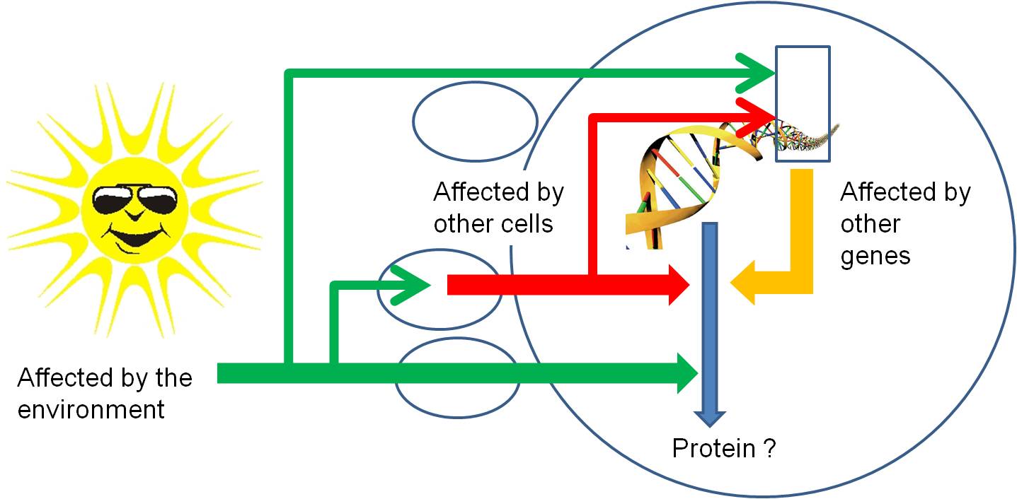
So, through all of these mechanisms,
protein synthesis can be stopped or stimulated, and the product can be modified.
Again, all of these regulatory pathways can be affected by environmental factors
or the proteins or mi/si-RNA's produced by other genes. So, gene activity is
affected by other things happening in the cell (turning other genes on and off)
, in other cells of the organism (through the production of hormones that act
as signal transducers), or environmental factors outside of the organism acting
directly on this gene, on other genes in this cell, or on other cells..
Cell division is the process of producing
two functional 'daughter' cells from one ancestral 'parental' cell. In order
for both of the daughter cells to have the full functional repertoire of the
original parental cell, they must be able to make the full complement of proteins
that the parent cell makes. In order for this to happen, they must both receive
the full complement of genetic information (DNA) in the parental cell. Hmmm....
how can they BOTH get the FULL COMPLEMENT of genetic information in the parental
cell? Well, in order for this to happen, the parental cell must duplicate its
DNA prior to cell division. This process of DNA replication produces two full
complements of genetic information. Then, this genetic information must be divided
evenly, in an organized manner, to insure that both daughter cells get the complete
complement of information (and not a duplication of some information or an omission
of other information). Cells that receive an incomplete complement of genetic
information will not be able to make all the proteins the parental cell made,
and may not be able to survive. So, again, DNA replication and the process of
mitosis are of great selective, adaptive value. Only cells that replicate and
divide their genetic information evenly, with only minor errors or inconsistencies,
will be likely to survive. These survivors will pass on the tendancy to replicate
and divide their genetic information evenly, as well. So, there is very strong
selection ( a very large selective advantage) for correct DNA replication and
equal chromosomal allocation during mitosis.
These processes of DNA replication
and mitosis are only two stages in the life of a cell. To place them in context,
it's useful to consider the full life of a cell, from it's production by the
division of its parental cell through to its own division.
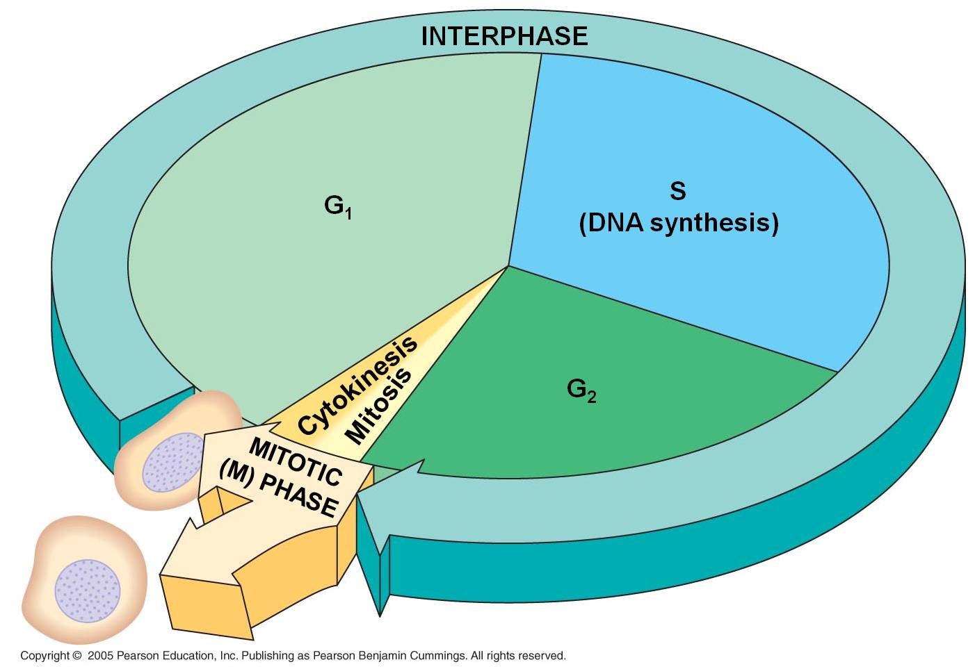 D.
Mitosis and the Cell Cycle
1. Interphase
- the 'interval' between divisions
a. G1
Our cell's life begins. That's sort
of a funny way to put it, because it seems to suggest that it is something new;
yet all of its constituents were part of the original parental cell. It is more
truly "1/2 an old cell with a full complement of DNA". Nevertheless,
it is an independent entity. In most protists, binary fission of the mitochondria
and chloroplasts occurs concurrently with the division of the nucleus during
mitosis, so the daughter cells have 'new' organelles, too. But in most multicellular
organisms, the allocation of organelles is largely a random process based on
how they are distributed in the cytoplasm during division. Then, the organelles
divide and 'repopulate' each daughter cell in G1.
The cell is roughly 1/2 the size
of the original parental cell. To grow to its appropriate size, it must synthesize
new biological molecules - and that means making the enzymes that will catalyze
those reactions. So, the DNA unwinds to the 'beads on a string' level, and the
genes between histones are available for transcription. When the DNA is unwound
('diffuse'), separate chromosomes cannot be seen with a light microscope. Rather,
the nucleus stains a uniform color except for one or several dark regions called
'nucleoli' (singular = nucleolus). These are areas were large amounts of r-RNA
are being synthesized and complexed with ribosomal proteins into functional
ribosomes. The ribosomes are exported from the nucleus to the cytoplasm, where
they will anchor to endoplasmic reticulum or the cytoskeleton.
Indeed, the G1 phase of a cell's
life is the most metabolically active period of it's life. It is growing in
size, and producing the proteins appropriate for its tissue type. Most cells
in multicellular organisms specialize during this period. Cells with very specific
structural adaptations to their specialized tissue type - like neurons with
long axons and muscle cells crammed with linear microfilaments - often remain
stalled in this stage after they become specialized; they do not divide again.
In this case, this stalled 'permanent' G1 phase is referred to a G0 ("G-nought').
 b.
S
b.
S
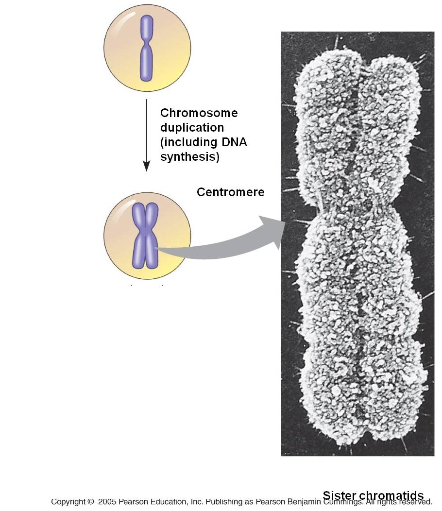
The S phase of the cell cycle is
when DNA replication occurs. The chromosomes are diffuse during this stage,
as well, so the enzymes (DNA polymerases) that replicate the DNA can access
the helices. Each double helix is separated, and the single strands are used
as templates for the formation of new helices on each template - changing one
double helix into two. Terminology becomes a bit ambiguous here. A DNA double
helix is equivalent to a "chromatid". A chromosome may have one chromatid
(in its unreplicated form) or two chromatids (in its replicated form). DNA replication
is a rather complicated process described in more detail below. The transition
from the G1 to the S phase is a very critical stage in a cell's life cycle,
signalling the cell's progression towards division. In eukaryotes it is called
a 'restriction point'. Once the S phase begins, the cell will proceed through
to mitosis. This transition is orchestrated by a complex interplay of transcription
factors that regulate the activity of "cell division cycle genes".
These genes produce cyclin proteins that vary in concentration through the cell
cycle. They bind with 'cyclin-dependent kinases' and these cdk-cyclin complexes
activate transcription factors that initiate the next phase of the cell cycle.
c. G2
After DNA replication is complete
the cell goes through another rapid period of growth in preparation for mitosis.
The DNA is checked again for damage caused and errors made during DNA replication.
Once again, p53 inhibits the transition to the mitotic phase, providing time
for this repair to take place. In cancer cells with mutations in p53, the G2
phase may be nearly eliminated, with the cell proceeding directly from DNA replication
to mitosis. CDK's bind to new cyclins, and these complexes active a different
set of proteins that initiate mitosis.
2. Mitosis
The process of mitosis can be summarized
as follows: the chromosomes condense, making it easier to divy them up evenly.
The replicated chromsomes are aligned in the middle of the cell by cytoskeletal
fibers. Each chromosome consistes of two identical double helices, called chromatids.
During the process of mitosis, these chromatids separate from each other, and
one double-helix from each chromosome is pulled to each end of the cell. The
membrane and cytoplasm are divided and the nuclear membrane reforms around the
chromosomes in each daughter cell. We will look at this process in more detail,
below.
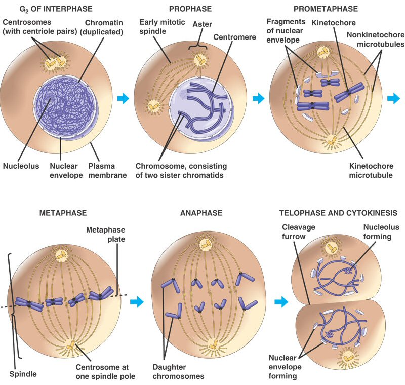
Mitosis is a continuous process of
chromosome condensation, chromatid separation, and cytoplasmic division. This
process is punctuated by particular events that are used to demarcate specific
stages. This process was first described by Walther Flemming in 1878, we he
developed new dyes and saw 'colored bodies' (chromo-somes) condensing and changing
position in dividing cells. He also coined the term 'mitosis' - the greek word
for thread - in honor of these thread-like structures.
1. Prophase: The transition from G2 to Prophase of Mitosis is marked by the condensation
of chromosomes.
2. Prometaphase: The chromosomes continue to condense, and the nuclear membrane disassembles.
The microfibers of the spindle apparatus attach to the kinetochores on the replicated
chromsomes.
3. Metaphase: The spindle aranges the chromosomes in the middle of the cell.
4. Anaphase: The proteins gluing sister chromatids together are metabolized, and the sister
chromatids are pulled by their spindle fibers to opposite poles of the cell.
It is important to appreciate that these separated chromatids (now individual,
unreplicated chromosomes) are idnetical to one another and identical to the
orignial parental chromosome (aside from unrepaired mutations).
5. Telophase: The cell continues to elongate, with a concentrated set of chromosomes at each
end. Nuclear membranes reform around each set of chromosomes, and the chromosomes
begin to decondense.
6. Cytokinesis: Cytokinesis is sometimes
considered a part of telophase. In this stage, the cytoplasm divides. In animal
cells, the membrane constricts along the cell's equator, causing a depression
or cleavage around the mid-line of the cell. This cleavage deepens until the
cells are pinched apart. In plants, vesicles from the golgi coalesce in the
middle of the cell, expanding to form a partition that divides the cell and
acts as a template for the deposition of lignin and cellulose that will form
the new cell wall between the cells.
As a consequence of this process,
two cells are produced from one parental cell, each having a complete complement
of genetic information - a copy of each original chromosome that was present
in the parental cell. Each of these cells now begins the G1 phase of interphase.
STUDY QUESTIONS:
1) Diagram the
parts of an RNA nucleotide.
2) Show
how two nucleotides are linked together by dehydration synthesis reactions.
3) Why does the
purine - pyrimidine structure relate to the complementary nature of double-stranded
DNA?
4) Draw
a DNA double helix, showing three base pairs and the antiparallel nature of
the helices.
5) Describe
the higher levels of eukaryotic chromosome structure, including the terms nucleosome
and solenoid.
6) What 'cues' determine where
transcription will start and stop?
7) What are introns and exons, and how are they processed?
8) Describe
translation: what is read, what is produced, and how is the 'genetic code' involved?.
9) How is
a polypeptide modified after translation to become a functional protein?
10) The process
of protein synthesis and the universal genetic code provides one of the dramatic
pieces of evidence of a common ancestry to all life. Explain why.
11) Draw
the cell cycle, labeling each stage and highlighting the main event in each
stage.
12) Draw a
chromsome before and after replication; use the terms chromosome and chromatid.
13) Draw a
cell, 2n = 6, and show each of the stages of mitosis. Write a brief description
of the events of each stage.
 Much of the energy harvested by a cell is used to make proteins. However, in order to understand protein synthesis, we must first describe the structure of DNA. DNA is the 'recipe' for proteins, so we need to take a little detour on our jounrey of how a cell works to describe the structure of DNA and chromosomes. DNA
is the genetic material in all forms of life (eubacteria, archaea, protists,
plants, fungi, and animals). Those quasi-living viruses vary in their genetic
material. Some have double-stranded DNA (ds-DNA) like living systems, while
others have ss-DNA, ss-RNA, and ds-RNA. RNA performs a wide array of functions
in living systems. Many of these functions have only been discovered in the
last few years.
  A.
DNA and RNA Structure
 -
Pentose (5 carbon) sugar: either ribose (RNA) or deoxyribose
(DNA). The carbons are numbered clockwise. The difference between the sugars
is that ribose has an -OH group on the 2' carbon, whereas deoxyriboes has
only 2 H groups and thus is "deoxygenated" relative to ribose. BOTH
sugars have an -OH group on the 3' carbon, which will be involved in binding.
The 5' carbon is a sidegroup off the ring.
 -
The third component of a nucleotide is a phosphate group,
which is attached to the 5' carbon of the sugar. When a nucleotide is incorporated
into a chain, it has a single phosphate group. However, nucleotides can occur
that have two or three phosphate groups (dinucleotides and trinucleotides).
ADP and ATP are important examples of these types of molecules. In fact, the
precursors of incorporated nucleotides are trinucleotides. When two phosphates
are cleaved, energy is released that can be used to add the remaining monophosphate
nucleotide to the nucleic acid chain.
 3.
Most DNA exists as a 'double helix' (ds-DNA) containing two linear nucleic acid
chains.
 1.
Overview
 a. The message is on one strand of the
double helix - the sense strand: The DNA double helix is composed
of two anti-parallel complementary strands of DNA. Only one strand in a coding
region ("gene") is read; this strand carries a meaningful recipe that
"makes sense". This is called the "sense" strand. The other
strand, limited by complementarity, is not a meaningful message - it is the
"non-sense" (or "anti-sense") strand. Think about it this
way. Given a meaningful message of "C-A-T" (a small furry mammal),
the complementary strand is limited to the meaningless sequence of "G-T-A"
(????...). As the meaningful sequence gets longer, it is even LESS likely that,
just by chance, the complementary strand would be meaningful, too. Again, in
all eukaryotic genes and in some rare prokaryotic ones, there will be non-functional
"introns" interspersed throughout the meaningful message. The meaningful
parts are called "exons". The process of transcription is continuous,
so introns and exons get transcribed and these regions - if present in the DNA
- will also be present in the RNA product.
 2.
Transcript Processing:
 a.
m-RNA attaches to the ribosome at the 5' end. The ribosome has two reactive sites. The RNA
moves through the ribosome until a specific sequence of nucleotides, AUG, is
positioned in the first site. Three base sequence in the m-RNA are called 'codons'.
This specific codon AUG, which starts the process of translation, is called
the 'start codon'. All proteins made by all life forms initially begin with
methionine, and use the codon AUG..
 1.
Regulation of Transcription:
 D.
Mitosis and the Cell Cycle
 b.
S
b.
S